A protein-structure record is a model built from experimental data. It is not a video of a reaction, a measurement in a person or a description of a retail kava product. The RCSB Protein Data Bank entry 6OP5 provides a useful example of how to read this evidence carefully. [1]
What the 6OP5 record contains
RCSB identifies 6OP5 as an X-ray diffraction structure of Piper methysticum styrylpyrone synthase 1 with p-coumaroyl-CoA, reported at 2.70 Å resolution. The enzyme comes from the kava plant; the record lists Escherichia coli BL21(DE3) as the expression system used to produce the protein for the experiment. Those are different biological roles and should not be collapsed into one claim. [1]
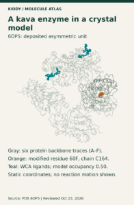
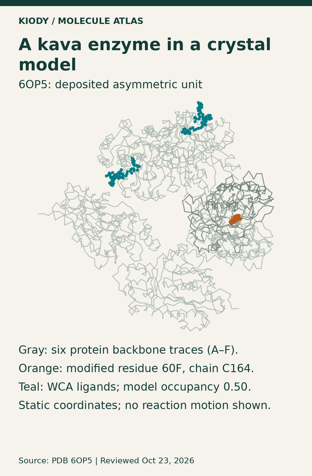
The deposited asymmetric unit contains six modeled protein chains. RCSB separately lists author-assigned biological assemblies as two-chain units. The six-chain drawing below therefore shows the deposited asymmetric unit, not a claim that the working enzyme is a six-subunit assembly. [1]
Reading the overview graphic
Gray lines follow modeled alpha-carbon positions in chains A–F. Orange marks modified residue 60F at chain C, residue 164. Teal marks the separately modeled p-coumaroyl-CoA ligands in chains A and B. The deposited atom-occupancy value for those displayed ligand atoms is 0.50.
Occupancy is a crystallographic model field. It is not a concentration, dose, binding-strength measurement or safety rating. Missing sequence stretches are left as gaps, and this rendering does not reconstruct electron density or show reaction motion.
A closer look at modified residue 60F
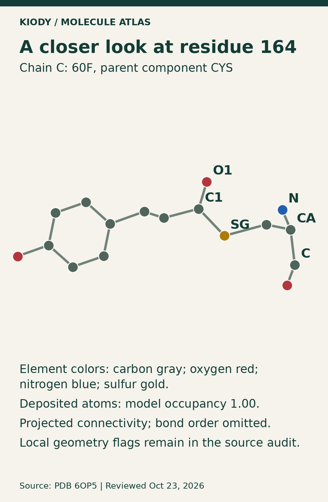
The second graphic projects the 17 atoms for 60F C164 that are present in the deposited coordinate model. Connections follow the deposited component-bond table only where both atoms are present. Bond multiplicities are intentionally omitted, so the drawing is not a complete chemical formula diagram.
The RCSB chemical-component record identifies 60F as an L-peptide-linking component with formula C12H13NO4S. The overview keeps the modified protein residue separate from the WCA ligand. [2]
The wwPDB validation report provides quality context for the deposited model, including residue-level geometry and electron-density fit. These graphics do not refit the structure or remove the local validation flags recorded in the source audit. [3]
What these graphics do—and do not—establish
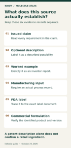
They show selected information from one deposited, static protein model. They do not establish human pharmacology, clinical effects, treatment, equivalence, safety, a manufacturing process or the formulation of any retail product. They also do not establish that every kava preparation contains the modeled enzyme or ligand complex.
Sources
[1] RCSB PDB, 6OP5: https://www.rcsb.org/structure/6OP5
[2] RCSB PDB, chemical component 60F: https://www.rcsb.org/ligand/60F
[3] wwPDB, full validation report: https://files.rcsb.org/validation/view/6op5_full_validation.pdf
[4] Deposited coordinates: https://files.rcsb.org/download/6OP5.cif
Reviewed October 23, 2026. Educational content for adults 21+.